On October 4, 2019 Cellular Imaging Facility Core manager Doug Cromey, MS was the invited speaker for a half-day workshop at Virginia Polytechnic Institute and State University entitled Introduction to Scientific Digital Images: Avoiding Twisted Pixels. Full Story: http://swehsc.pharmacy.arizona.edu/digital-imaging-workshop-VT Read more »
Dancing Atoms Reveal Potential Capabilities of Materials
An important part of Peter Crozier’s job involves watching dances. He views these intricate performances through state-of-the-art, high-powered microscopes because the dancers are atoms. Crozier is a materials scientist at Arizona State University who studies how the underlying principles of […] Read more »
Imaging at the Speed of Life

To study the swiftness of biology – the protein chemistry behind every life function – scientists need to see molecules changing and interacting in unimaginably rapid time increments: trillionths of a second or shorter. Imaging equipment with that kind of […] Read more »
UA to add several new high-end Microscopes
Senior Vice President for Research Kimberly Espy has approved funding to move forward with the purchase of three high-end optical microscopes from Carl Zeiss Microscopy, LLC. Zeiss had the winning proposal in a competitive RFP that was conducted this summer. […] Read more »
New Cryo-EM at the Southwest Regional Center
The hottest technology in all of science will soon bring a new coolness factor to world-class Arizona State University research. The coolest new way to take a near-atomic resolution snapshot of life at work is cryo-Electron Microscopy (cryo-EM) – lauded […] Read more »
USA Today: Increasingly, dispensaries and patients are turning to laboratories to evaluate plants
An article published in the Arizona Republic on January 6th, was picked up by USA Today about private research groups screening medical marijuana from dispensaries to screen for molds or pesticides. In addition to testing for contaminants that may cause illness in those with a weakened immune system, the […] Read more »
Ever hear of a Foldscope?
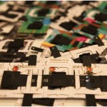
Stanford researcher Manu Prakash developed a microscope that can fit into your pocket! It’s a 50-cent print-and-fold paper microscope that uses a watch battery, LED and a few optical units that can magnify objects up to 2,000 times. The original […] Read more »
ASU 2015 Winter School on High Resolution Electron Microscopy
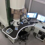
The LeRoy Erying Center for Solid State Science again offered their Electron Microscopy School during January 2015 to provide advanced training to scientists who use TEM microscopes for material science studies. The course demonstrated environmental electron microscopy, focused ion beam […] Read more »
Carbon under pressure exhibits some interesting traits
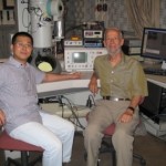
High pressures and temperatures cause materials to exhibit unusual properties, some of which can be special. Understanding such new properties is important for developing new materials for desired industrial uses and also for understanding the interior of Earth, where everything […] Read more »
New grant advances ASU microscopy imaging initiative
Peering through a homemade instrument – toy-like by today’s standards – the Dutch tradesman Antony van Leeuwenhoek (1632-1723) first observed a dizzying menagerie of lifeforms, invisible to the naked eye. Since then, scientists have steadily refined the field of microscopy, […] Read more »